Oogenesis
Oogenesis—the differentiation of the ovum—differs from spermatogenesis in several ways. Whereas the gamete formed by spermatogenesis is essentially a motile nucleus, the gamete formed by oogenesis contains all the materials needed to initiate and maintain metabolism and development. Therefore, in addition to forming a haploid nucleus, oogenesis also builds up a store of cytoplasmic enzymes, mRNAs, organelles, and metabolic substrates. While the sperm becomes differentiated for motility, the egg develops a remarkably complex cytoplasm.
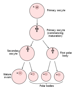
The mechanisms of oogenesis vary among species more than those of spermatogenesis. This difference should not be surprising, since patterns of reproduction vary so greatly among species. In some species, such as sea urchins and frogs, the female routinely produces hundreds or thousands of eggs at a time, whereas in other species, such as humans and most mammals, only a few eggs are produced during the lifetime of an individual. In those species that produce thousands of ova, the oogonia are self-renewing stem cells that endure for the lifetime of the organism. In those species that produce fewer eggs, the oogonia divide to form a limited number of egg precursor cells. In the human embryo, the thousand or so oogonia divide rapidly from the second to the seventh month of gestation to form roughly 7 million germ cells . After the seventh month of embryonic development, however, the number of germ cells drops precipitously. Most oogonia die during this period, while the remaining oogonia enter the first meiotic division . These latter cells, called the primary oocytes, progress through the first meiotic prophase until the diplotene stage, at which point they are maintained until puberty. With the onset of adolescence, groups of oocytes periodically resume meiosis. Thus, in the human female, the first part of meiosis begins in the embryo, and the signal to resume meiosis is not given until roughly 12 years later. In fact, some oocytes are maintained in meiotic prophase for nearly 50 years. As indicates, primary oocytes continue to die even after birth. Of the millions of primary oocytes present at birth, only about 400 mature during a woman's lifetime.
The mechanisms of oogenesis vary among species more than those of spermatogenesis. This difference should not be surprising, since patterns of reproduction vary so greatly among species. In some species, such as sea urchins and frogs, the female routinely produces hundreds or thousands of eggs at a time, whereas in other species, such as humans and most mammals, only a few eggs are produced during the lifetime of an individual. In those species that produce thousands of ova, the oogonia are self-renewing stem cells that endure for the lifetime of the organism. In those species that produce fewer eggs, the oogonia divide to form a limited number of egg precursor cells. In the human embryo, the thousand or so oogonia divide rapidly from the second to the seventh month of gestation to form roughly 7 million germ cells . After the seventh month of embryonic development, however, the number of germ cells drops precipitously. Most oogonia die during this period, while the remaining oogonia enter the first meiotic division . These latter cells, called the primary oocytes, progress through the first meiotic prophase until the diplotene stage, at which point they are maintained until puberty. With the onset of adolescence, groups of oocytes periodically resume meiosis. Thus, in the human female, the first part of meiosis begins in the embryo, and the signal to resume meiosis is not given until roughly 12 years later. In fact, some oocytes are maintained in meiotic prophase for nearly 50 years. As indicates, primary oocytes continue to die even after birth. Of the millions of primary oocytes present at birth, only about 400 mature during a woman's lifetime.
